


 醫學研究 學術文章 Ablative Chemoembolization for Hepatocellular Carcinoma: A Prospective Phase I Case-Control Comparison with Conventional Chemoembolization. Yu SCH, Chan SL, Lee KF, Hui JWY, Hui EP, Chu CM,et al. Radiology. 2018 Apr;287(1):340-348. doi: 10.1148/radiol.2017170154. Epub 2017 Dec 22. PMID: 29272212.
肝癌 Liver cancer
Ablative Chemoembolization for Hepatocellular Carcinoma: A Prospective Phase I Case-Control Comparison with Conventional Chemoembolization.
Yu SCH, Chan SL, Lee KF, Hui JWY, Hui EP, Chu CM, Chan AW, Cheung S, Li L, Wong J, Yeo WMM. Radiology. 2018 Apr;287(1):340-348. doi: 10.1148/radiol.2017170154. Epub 2017 Dec 22. PMID: 29272212.
Abstract
Purpose: To evaluate the feasibility, safety, and treatment effectiveness of ablative chemoembolization (ACE) in the treatment of hepatocellular carcinoma (HCC) and compare with a similar patient cohort who underwent conventional transarterial chemoembolization (cTACE).
Materials and Methods: This was a prospective phase I nonrandomized study conducted between March 2013 and October 2016 in accordance to the Declaration of Helsinki and Declaration Good Clinical Practice with written informed consent. There were 36 men and eight women (median age, 64 years [interquartile range, 58-74] and 74.5 years [interquartile range, 70-80], respectively). The primary end points were treatment safety and tumor response. The secondary end points were time to progression, progression-free survival, conversion to partial hepatectomy, and viable HCC within the tumor specimen. The end points of the study group (n = 22) were compared with those of a case-matched control group (n = 22) of patients who underwent conventional cTACE during the same period by using a Pearson χ2 test.
Results: Treatment with ACE was successfully completed in all patients without adverse effects. The complete response (CR) rates by patient or by tumor were both 100%. The median time to progression and median progression-free survival were significantly longer in the study group than in the control group (both were 28 months vs 10 months, respectively; P < .001). The number of patient conversions to hepatectomy was seven for ACE and three for cTACE. In the tumor specimens, viable tumor was found in two of eight specimens that underwent ACE and three of three that underwent cTACE.
Conclusion: ACE is a feasible, safe, and well-tolerated treatment for patients with HCC; it is highly effective and may be more effective than cTACE in achieving CR.
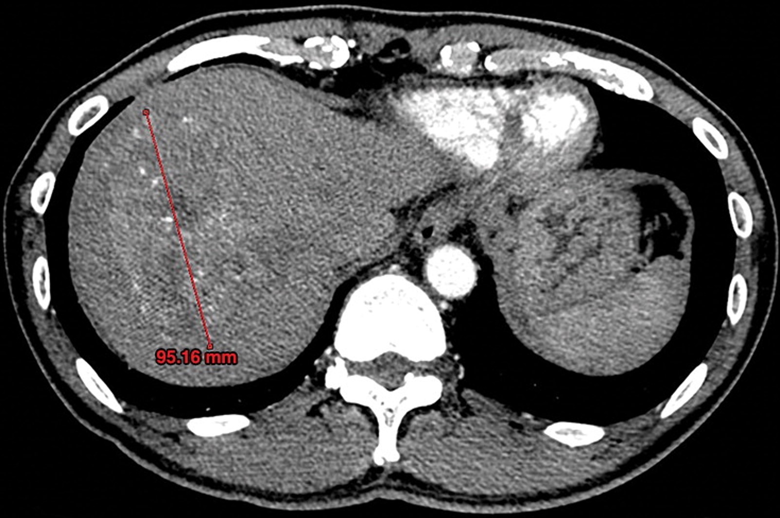
Figure 2a: (a) In a 56 year-old male patient, arterial-phase CT showed a 9.5-cm tumor with contrast enhancement typical of HCC in segment VII and VIII. (b) Digital subtraction angiography showed a well-defined tumor of hypervascularity typical of massive-expansive HCC. (c) Immediately after ACE, in which 20 mL of lipiodol-ethanol-cisplatin was administered, radiography showed lipiodol accumulation within the intratumoral and peritumoral vasculature. (d) Digital subtraction angiography performed at 2 months after the first treatment showed no residual intratumoral vascularity. (e) CT at 3 months after the first treatment showed no evidence of residual enhancing tissue and size reduction to 6.8 cm. (f) The patient underwent right hepatectomy 7 months after undergoing ACE, and this cross-section of the surgical specimen shows the tumor. (g) Histopathologic assessment of the surgical specimen by using hematoxylin-and-eosin stain showed no evidence of viable residual HCC within the tumor (black arrows), the capsule (white arrows), and liver tissue (*).
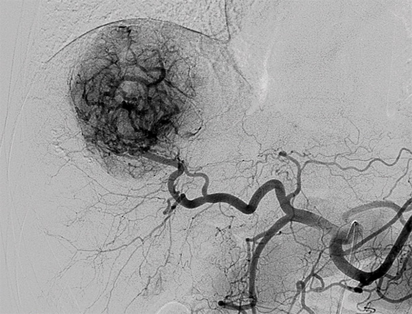
Figure 2b: (a) In a 56 year-old male patient, arterial-phase CT showed a 9.5-cm tumor with contrast enhancement typical of HCC in segment VII and VIII. (b) Digital subtraction angiography showed a well-defined tumor of hypervascularity typical of massive-expansive HCC. (c) Immediately after ACE, in which 20 mL of lipiodol-ethanol-cisplatin was administered, radiography showed lipiodol accumulation within the intratumoral and peritumoral vasculature. (d) Digital subtraction angiography performed at 2 months after the first treatment showed no residual intratumoral vascularity. (e) CT at 3 months after the first treatment showed no evidence of residual enhancing tissue and size reduction to 6.8 cm. (f) The patient underwent right hepatectomy 7 months after undergoing ACE, and this cross-section of the surgical specimen shows the tumor. (g) Histopathologic assessment of the surgical specimen by using hematoxylin-and-eosin stain showed no evidence of viable residual HCC within the tumor (black arrows), the capsule (white arrows), and liver tissue (*).
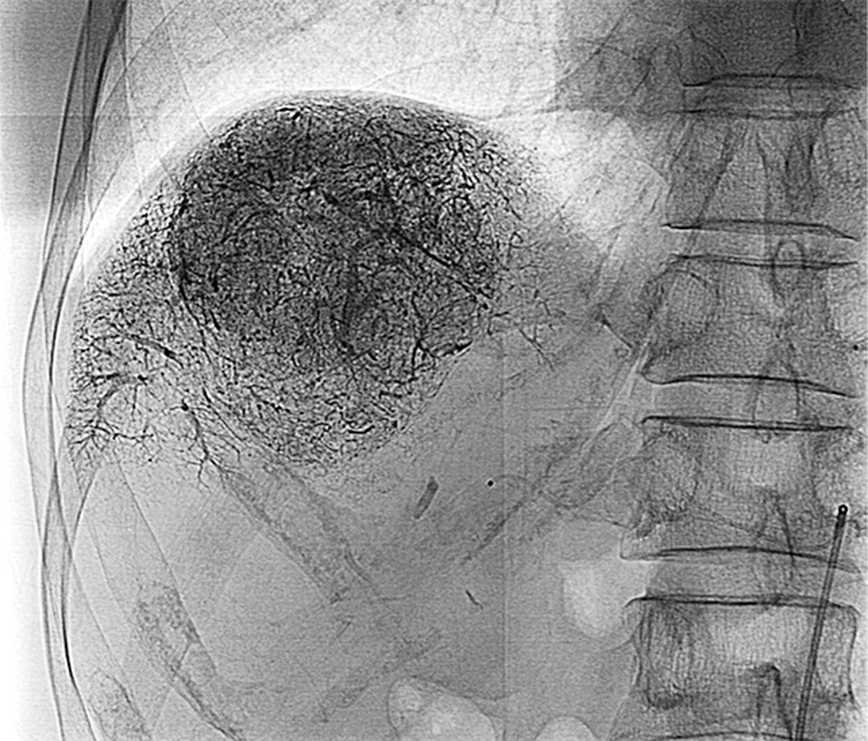
Figure 2c: (a) In a 56 year-old male patient, arterial-phase CT showed a 9.5-cm tumor with contrast enhancement typical of HCC in segment VII and VIII. (b) Digital subtraction angiography showed a well-defined tumor of hypervascularity typical of massive-expansive HCC. (c) Immediately after ACE, in which 20 mL of lipiodol-ethanol-cisplatin was administered, radiography showed lipiodol accumulation within the intratumoral and peritumoral vasculature. (d) Digital subtraction angiography performed at 2 months after the first treatment showed no residual intratumoral vascularity. (e) CT at 3 months after the first treatment showed no evidence of residual enhancing tissue and size reduction to 6.8 cm. (f) The patient underwent right hepatectomy 7 months after undergoing ACE, and this cross-section of the surgical specimen shows the tumor. (g) Histopathologic assessment of the surgical specimen by using hematoxylin-and-eosin stain showed no evidence of viable residual HCC within the tumor (black arrows), the capsule (white arrows), and liver tissue (*).
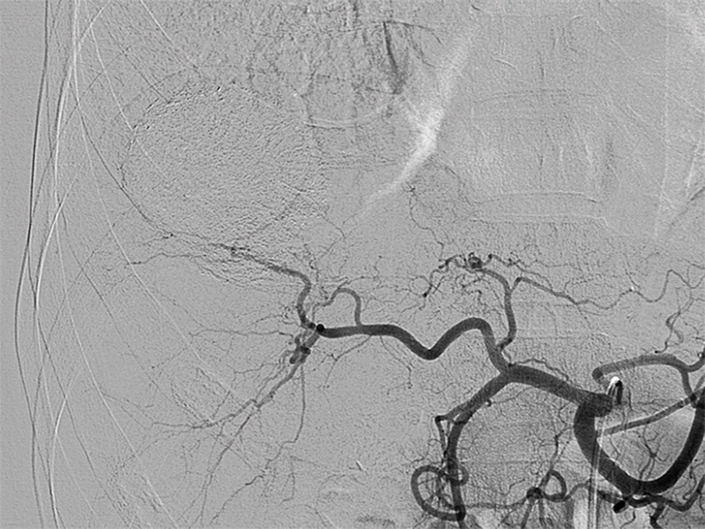
Figure 2d: (a) In a 56 year-old male patient, arterial-phase CT showed a 9.5-cm tumor with contrast enhancement typical of HCC in segment VII and VIII. (b) Digital subtraction angiography showed a well-defined tumor of hypervascularity typical of massive-expansive HCC. (c) Immediately after ACE, in which 20 mL of lipiodol-ethanol-cisplatin was administered, radiography showed lipiodol accumulation within the intratumoral and peritumoral vasculature. (d) Digital subtraction angiography performed at 2 months after the first treatment showed no residual intratumoral vascularity. (e) CT at 3 months after the first treatment showed no evidence of residual enhancing tissue and size reduction to 6.8 cm. (f) The patient underwent right hepatectomy 7 months after undergoing ACE, and this cross-section of the surgical specimen shows the tumor. (g) Histopathologic assessment of the surgical specimen by using hematoxylin-and-eosin stain showed no evidence of viable residual HCC within the tumor (black arrows), the capsule (white arrows), and liver tissue (*).
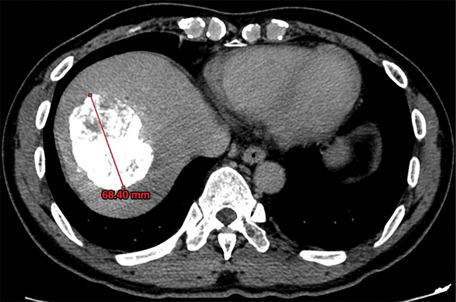
Figure 2e: (a) In a 56 year-old male patient, arterial-phase CT showed a 9.5-cm tumor with contrast enhancement typical of HCC in segment VII and VIII. (b) Digital subtraction angiography showed a well-defined tumor of hypervascularity typical of massive-expansive HCC. (c) Immediately after ACE, in which 20 mL of lipiodol-ethanol-cisplatin was administered, radiography showed lipiodol accumulation within the intratumoral and peritumoral vasculature. (d) Digital subtraction angiography performed at 2 months after the first treatment showed no residual intratumoral vascularity. (e) CT at 3 months after the first treatment showed no evidence of residual enhancing tissue and size reduction to 6.8 cm. (f) The patient underwent right hepatectomy 7 months after undergoing ACE, and this cross-section of the surgical specimen shows the tumor. (g) Histopathologic assessment of the surgical specimen by using hematoxylin-and-eosin stain showed no evidence of viable residual HCC within the tumor (black arrows), the capsule (white arrows), and liver tissue (*).
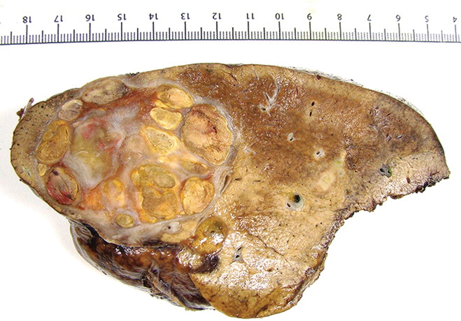
Figure 2f: (a) In a 56 year-old male patient, arterial-phase CT showed a 9.5-cm tumor with contrast enhancement typical of HCC in segment VII and VIII. (b) Digital subtraction angiography showed a well-defined tumor of hypervascularity typical of massive-expansive HCC. (c) Immediately after ACE, in which 20 mL of lipiodol-ethanol-cisplatin was administered, radiography showed lipiodol accumulation within the intratumoral and peritumoral vasculature. (d) Digital subtraction angiography performed at 2 months after the first treatment showed no residual intratumoral vascularity. (e) CT at 3 months after the first treatment showed no evidence of residual enhancing tissue and size reduction to 6.8 cm. (f) The patient underwent right hepatectomy 7 months after undergoing ACE, and this cross-section of the surgical specimen shows the tumor. (g) Histopathologic assessment of the surgical specimen by using hematoxylin-and-eosin stain showed no evidence of viable residual HCC within the tumor (black arrows), the capsule (white arrows), and liver tissue (*).
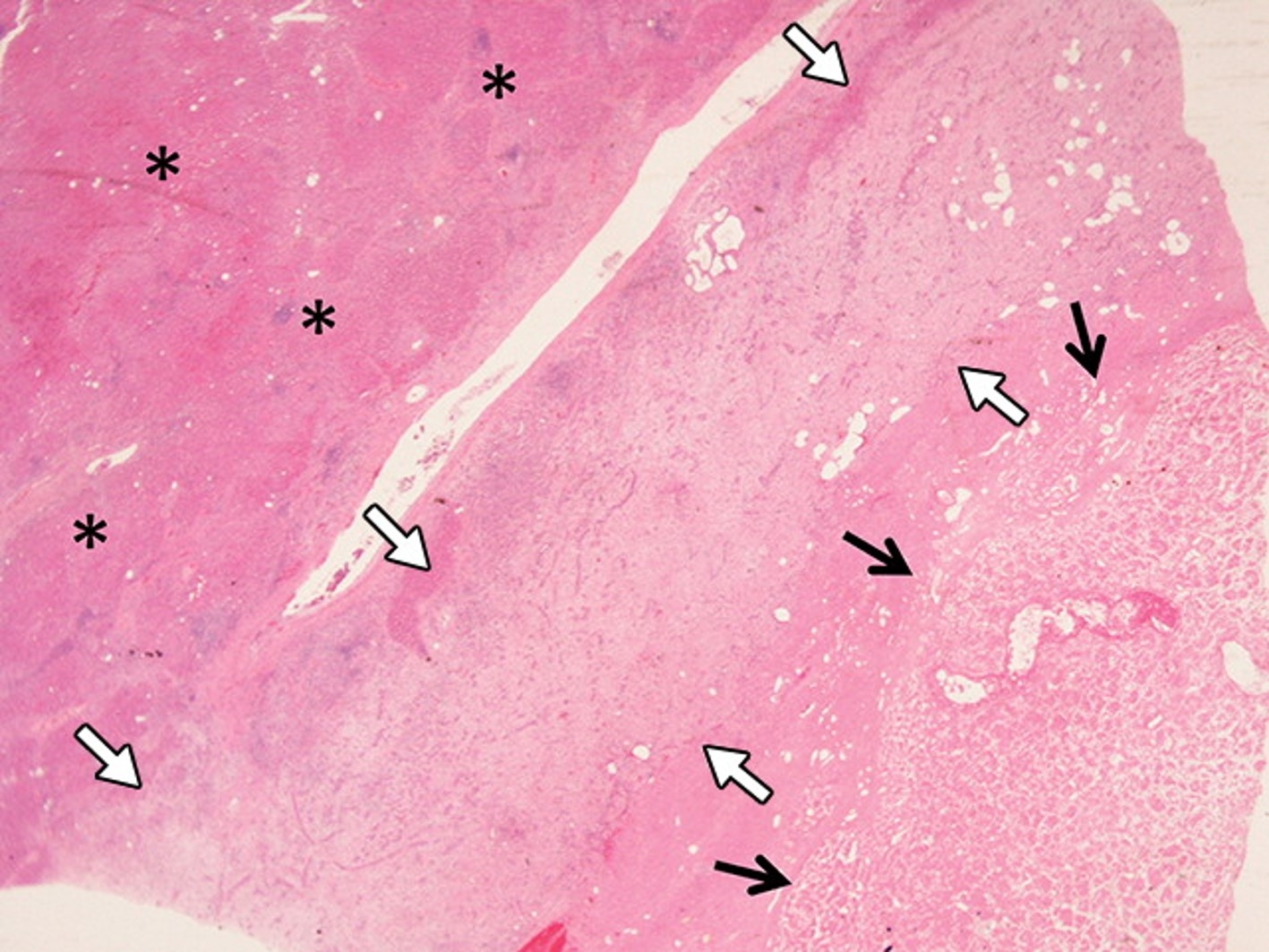
Figure 2g: (a) In a 56 year-old male patient, arterial-phase CT showed a 9.5-cm tumor with contrast enhancement typical of HCC in segment VII and VIII. (b) Digital subtraction angiography showed a well-defined tumor of hypervascularity typical of massive-expansive HCC. (c) Immediately after ACE, in which 20 mL of lipiodol-ethanol-cisplatin was administered, radiography showed lipiodol accumulation within the intratumoral and peritumoral vasculature. (d) Digital subtraction angiography performed at 2 months after the first treatment showed no residual intratumoral vascularity. (e) CT at 3 months after the first treatment showed no evidence of residual enhancing tissue and size reduction to 6.8 cm. (f) The patient underwent right hepatectomy 7 months after undergoing ACE, and this cross-section of the surgical specimen shows the tumor. (g) Histopathologic assessment of the surgical specimen by using hematoxylin-and-eosin stain showed no evidence of viable residual HCC within the tumor (black arrows), the capsule (white arrows), and liver tissue (*).
Citation: