


 醫學研究 學術文章 Transarterial Ethanol Ablation for Unresectable Hepatocellular Carcinoma: Analysis of Clinical and Tumor Outcomes. Yu SC, Hui EP, Tang P, Chan SK, Chu CC, Hui JW, et al. J Vasc Interv Radiol. 2016 May;27(5):639-49. doi: 10.1016/j.jvir.2015.11.032. Epub 2016 Jan 20. PMID: 26803574.
肝癌 Liver cancer
Transarterial Ethanol Ablation for Unresectable Hepatocellular Carcinoma: Analysis of Clinical and Tumor Outcomes
Yu SC, Hui EP, Tang P, Chan SK, Chu CC, Hui JW, Chan SL, Lee KF, Lai PB, Yeo W. J Vasc Interv Radiol. 2016 May;27(5):639-49. doi: 10.1016/j.jvir.2015.11.032. Epub 2016 Jan 20. PMID: 26803574.
Abstract
Purpose: To evaluate survival, tumor response, and treatment toxicity of transarterial ethanol ablation (TEA) in patients with unresectable hepatocellular carcinoma (HCC).
Materials and Methods: This prospective study involved 186 patients (146 men and 40 women; median age, 65 y [interquartile range, 57–72.3 y]). Of 186 patients, 146 (78.5%) were hepatitis B virus carriers, 18 (9.7%) were hepatitis C virus carriers, 82 (44.1%) had tumors ≥ 5 cm, and 43 (23.1%) had multifocal tumors. Overall survival (OS), complete response (CR) by European Association for the Study of the Liver criteria, time to progression (TTP), progression-free survival (PFS), and treatment toxicities were evaluated. Univariate and multivariate analyses for prognostic factors of OS were performed.
Results: Median OS was 25.7 months (95% confidence interval [CI], 20.9–30.5) and varied significantly between Child-Pugh A and B (28.7 mo vs 13.4 mo, P < .001), and Barcelona Clinic Liver Cancer A and B or C (37.1 mo vs 17.7 mo, P = .001). P rognostic factors for longer OS were solitary tumor, tumor size < 5 cm, > 1 treatments, and CR of all tumors at 6 months. TTP was 9.1 months (95% CI, 6.9–11.3). PFS was 8.4 months (95% CI, 7.1–9.7). CR occurred in 69.1% (159/230) of lesions and 48.9% (88/180) of patients at 6 months. Any one symptom of the postembolization syndrome of grade 2 severity occurred in < 22% (41/186) of patients. No treatment-related hepatitis or death occurred within 30 days. Transient respiratory decompensation occurred in three patients (1.6% [3/186]), and alcoholic intoxication occurred in one patient (0.5% [1/186]).
Conclusion: TEA appears to be safe and effective for local control of HCC.
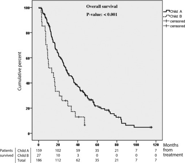
Figure 1. Kaplan-Meier curves of OS in subgroups classified according to the Child-Pugh (Child) cirrhosis classification.
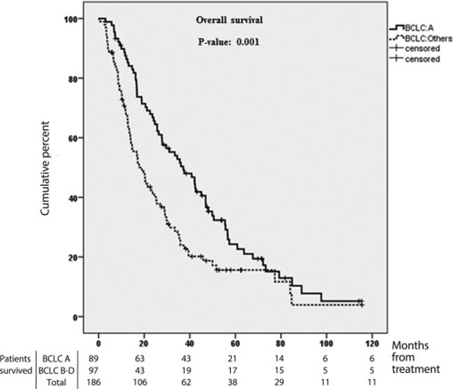
Figure 2. Kaplan-Meier curves of OS in subgroups classified according to the Barcelona Clinic Liver Cancer (BCLC) staging classification.
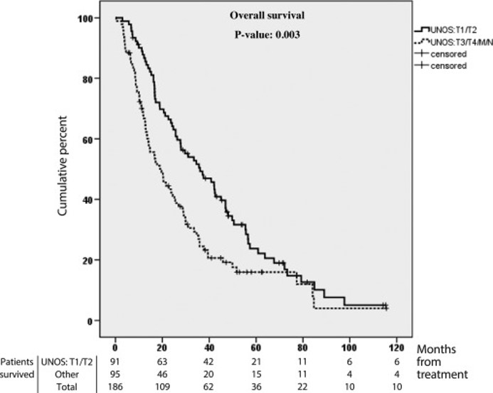
Figure 3. Kaplan-Meier curves of OS in subgroups classified according to the United Network for Organ Sharing (UNOS) staging classification.
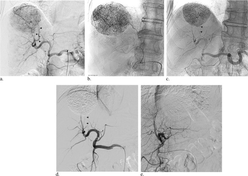
Figure 4. In a 51-year-old man with HBV-related HCC, a celiac axis arteriogram obtained with digital subtraction arteriography showed a massive expansive hypervascular tumor in segment VII/VIII, which was supplied by a hypertrophied feeding artery (arrowheads) (a) . At the completion of LEM administration through a microcatheter that was introduced up to the tumor boundary, the tumor vasculature and peritumoral portal venules were outlined with Lipiodol on radiography (b) . Arteriogram obtained at 2 months showed a markedly shrunken tumor with a well-defined margin and Lipiodol retention. The arrowheads mark the position of the feeding artery which is now not opacified (c) . Digital subtraction arteriography performed at two different projections showed occlusion of the feeding artery (arrowheads) and absence of intratumoral or peritumoral vascularity (d, e) .
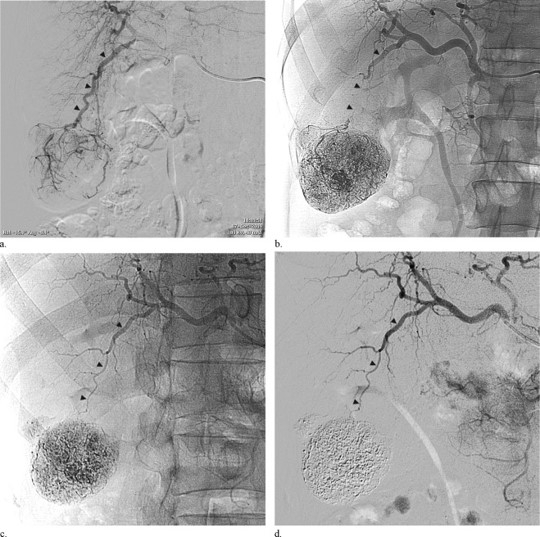
Figure 5. In a 64-year-old man with HBV-related HCC, a proper hepatic artery arteriogram obtained with digital subtraction arteriography showed a massive expansive hypervascular tumor in segment VI with a satellite lesion at its superolateral aspect, which was supplied by a hypertrophied feeding artery (arrowheads) (a) . A proper hepatic artery arteriogram obtained at the completion of LEM administration showed occlusion of the feeding artery (arrowheads) and Lipiodol deposition within the tumor, its satellite lesion, and peritumoral portal venules (b) . A proper hepatic artery arteriogram obtained at 2 months showed a markedly shrunken tumor with a well-defined margin and Lipiodol retention and a patent, normal-sized feeding artery (arrowheads) (c) . Digital subtraction arteriography showed patency and normal size of the feeding arterial branch (arrowheads) and absence of intratumoral or peritumoral vascularity (d) .
Citation: